By Eric Stann | MU News Bureau
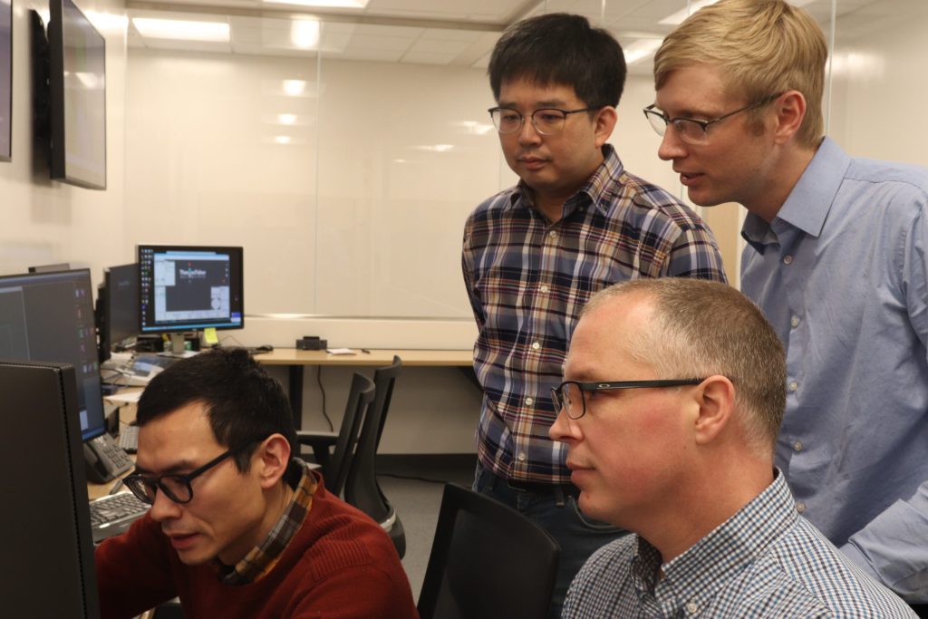
For more than 20 years, Matt Maschmann, an associate professor of mechanical and aerospace engineering at the University of Missouri, has worked with materials that require specialized technology — electron microscopes — to be seen by the human eye.
“When we deal with materials interacting on a nanoscale level, we can’t physically see the processes that are occurring, like the charging and discharging of a battery, for instance, without the help of an electron microscope,” Maschmann said.
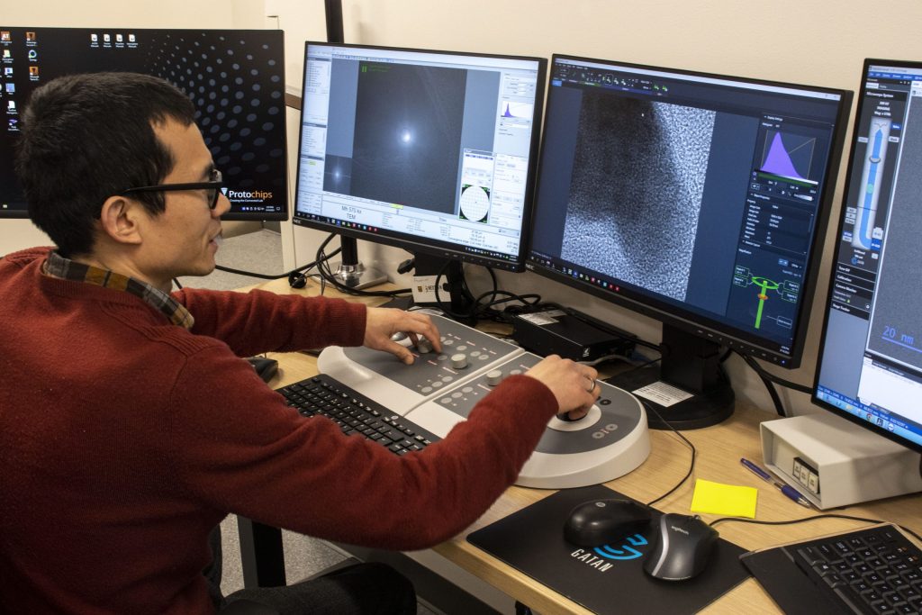
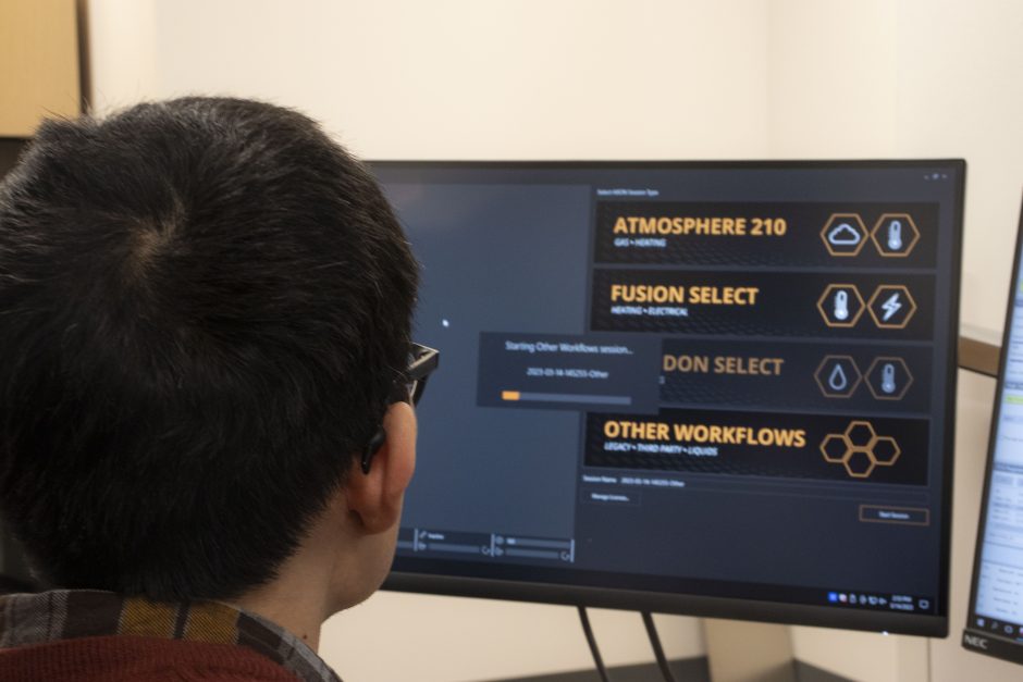
Now, with the support of a two-year, $800,000 grant from the National Science Foundation and an additional $300,000 from the university, Maschmann and a team of researchers at MU are purchasing new equipment from Protochips which will allow researchers to conduct scientific experiments while simultaneously viewing reactions as they happen in real time — under the lens of a Thermo Fisher Scientific Spectra 300 Transmission Electron Microscope (TEM), housed in the Electron Microscopy Core (EMC) facility located in the Roy Blunt NextGen Precision Health building.
“The TEM is capable of letting researchers visualize reactions and material changes with up to atomic resolution, which is amazing,” Maschmann said. “But conducting experiments within a TEM environment is challenging because only small amounts of material will fit into a TEM chamber, and the system operates in a high-vacuum environment. Traditionally, researchers acquire TEM images of what a material looks like before and after an experiment, which leaves them to make an educated hypotheses about why and how a material has changed.”
With the new equipment, Maschmann said researchers will be able to gain a greater understanding of what’s going on at the most basic level of material’s structure by seeing an entire experiment unfold in real time. He said they can also use the new equipment to conduct experiments in three different environments — liquid, gas or vacuum — within the electron microscope. These new capabilities will allow them to explore and observe how different materials react to different environmental stimuli, like heat or electricity.
“For instance, we will be able to examine the composition of nanomaterials all the way down to the atomic level,” said Maschmann, who became interested in doing scientific research by working as an undergraduate student with Hongbin “Bill” Ma, who is now one of his colleagues at MU. “We can obtain detailed measurements by seeing the atoms moving and can even determine what element the atoms are.”
The new equipment also includes active image stabilization, which prevents images from moving, blurring or going out of focus while researchers follow a reaction.
“If we didn’t have that [active image stabilization], it would be a big problem with these types of experiments,” Maschmann said. “For example, if we rapidly heat up a material, it’s going to expand and move. So, if our original field of view is only a few hundred nanometers, as soon as heat is applied our area of interest will move beyond our original viewing area. Therefore, we would likely miss the reaction that we intended to observe.”
There’s still much to be explored at this level of material science, and Maschmann is excited for the opportunities that this new equipment can provide to help advance scientific research across MU and beyond. The EMC facility is available to support scientific research by faculty from any of the four UM System universities, and that includes the new equipment for the TEM, he said.
The grant also highlights an interdisciplinary collaboration between the EMC and the MU Materials Science & Engineering Institute (MUMSI). The goal of MUMSI, which opened in 2021 as a partnership between the MU College of Engineering and the MU College of Arts and Science, is to advance collaboration around materials research and engineering education across the university. Maschmann is also the co-director of MUMSI.
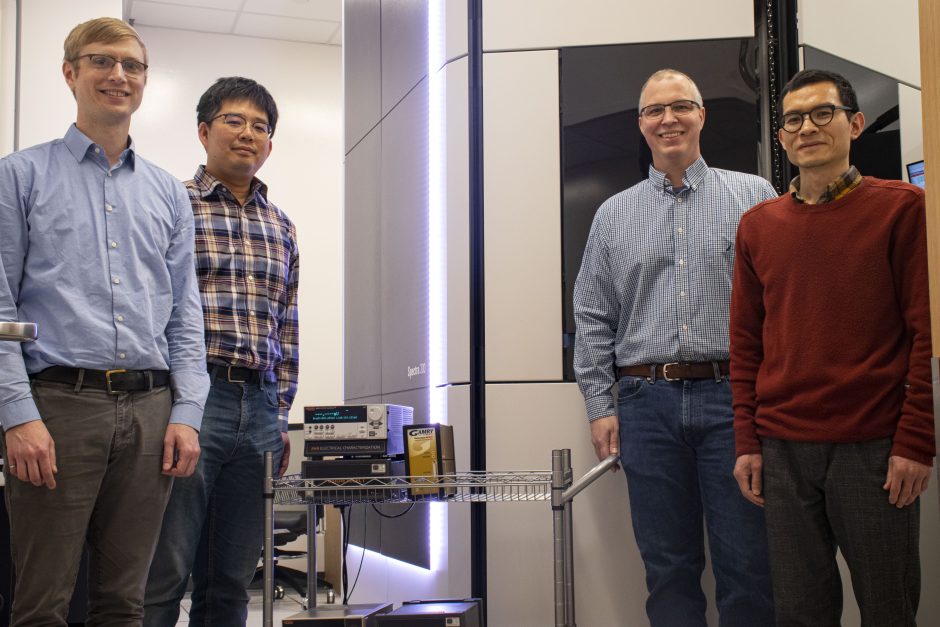
“MRI: Acquisition of Active Holders for in-situ and in-operando Transmission Electron Microscope Experiments,” was awarded by the National Science Foundation (2216026). Matthias J. Young, Xiaoqing He, Jaewon Lee and Maria Mills at MU are also involved with this project. The content is solely the responsibility of the authors and does not necessarily represent the official views of the funding agencies.